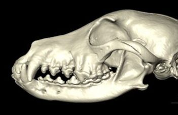
Comparing canine brains using 3D-endocast modelling

Reconstructing the brain based on the internal surface patterns of a skull is a well-known method among archeologists studying the brains of extinct species, e.g. dinosaurs, mammoths and even cavemen. One way to achieve a model of the brain is to fill the endocranial space with specific gradually hardening fluids, and after the solidifying of the material the bones can be removed to reveal the cast, which reflects the surface of the brain with high fidelity. These are called endocranial casts, or endocasts.
Nowadays, rather than utilising endocasts, invasive process diagnostic imaging techniques are highly preferred. Computer tomography (CT) is the best and easiest way to visualize the bones, with all of their external and internal structures and composition. Using CT is also advantageous in endocranial analysis, as researchers can create virtual endocasts based on the digitized skulls. This allows for a higher freedom during examinations, because these virtual models can be rotated, colorized, cut, and completed. They can be shown not only on mobile phones, tablets or computers, but with the help of 3D-printing technology one can replicate them in the requested number and scale as well.

Skull and endocast of a beagle. Video created by dr. Kálmán Czeibert
This digital endocasting technique was used by researchers from ELTE who scanned skulls from the collection of dr. Tibor Csörgő (ELTE, Department of Anatomy, Cell and Developmental Biology), which comprises almost 400 dog skulls from 152 canine breeds. “Due to the high resolution scanning, detailed 3D models could be created. This means that not only the main convolutions, that is, the cerebral gyri, can be recognized on the final model, but the location of the major blood vessels can also be seen on the surface.” – explains dr. Kálmán Czeibert, veterinarian and postdoctoral researcher at the Senior Family Dog Project, first author of the publication. “We compared the shape, volume and surface features of the endocasts from 28 specimens in our recent study in Frontiers in Veterinary Science. These models clearly show how the shortening of the facial part of the skull affected the morphology of the brain, distorting the olfactory bulb, which is pushed back and underneath the frontal lobe in certain breeds (like the French bulldog or pug), changing the ratio between the cerebral areas.”
In order to clarify how this change affects dogs’ olfactory and cognitive performance, further imaging and behavioral investigations are needed. The new collection is part of the Canine Brain and Tissue Bank at ELTE, funded by a European Research Council (ERC) grant. The bank will help researchers to link the brain with behaviour. “In some areas, where volume has diminished, neuronal density and the relationship with other brain regions may act to counteract these changes. To further examine these speculations, behavioural testing, diagnostic imaging (like CT, MR or EEG), and histological and molecular assessment will be needed.” – said dr. Enikő Kubinyi, senior researcher at ELTE, principal investigator of the research group.
“One of our primary aims was the widespread dissemination of scientific knowledge.’ – adds dr. Czeibert. “How dog skulls and brains vary is striking. For example, despite the 30-fold difference in body weight between a Neapolitan mastiff and a chihuahua, their endocasts’ volumes have only a 2-fold difference.” The researchers from ELTE plan to increase the number of skulls by digitizing more specimens for microevolutionary studies in the framework of an international cooperation.

The skull and endocast of a chihuahua and a Neapolitan mastiff. Image by dr. Kálmán Czeibert

A Neapolitan mastiff and a chihuahua, with their endocasts. Image created by dr. Kálmán Czeibert
Source of the dog photos: Wikimedia Commons
23 images in Dropbox:
https://www.dropbox.com/sh/hs68ennf6ygsma4/AAADlZGCsLsljCwUICqgHj8za?dl=0
Skulls and endocasts from a wolf and various breeds: American bulldog, Australian shepherd, beagle, border collie, borzoi, boxer, bull terrier, Cavalier King Charles spaniel, chihuahua, chow chow, doberman pinscher, English bulldog, English setter, French bulldog, German shepherd, komondor, Neapolitan mastiff, pug, rough collie.
Same images in Google Photos: https://photos.app.goo.gl/yRCLCQoYaNx4wWEH8
Same images in Google Drive (if the link does not work from the doc please copy it into your browser):
https://drive.google.com/drive/folders/1WzINnr1Y8J2Aoy3nnyHnSrC3MYEdbRSN?usp=sharing
Video: https://youtu.be/R7k-b-soVN4
Original study: Czeibert, K., Sommese, A., Petneházy, Ö., Csörgő, T., Kubinyi, E.: Digital endocasting in comparative canine brain morphology, Frontiers in Veterinary Science. 7. 2020. https://doi.org/10.3389/fvets.2020.565315
The research was supported by “Cognitive Ageing in Dogs” starting grant (EVOLOR 680040) of the European Research Council (ERC).

